

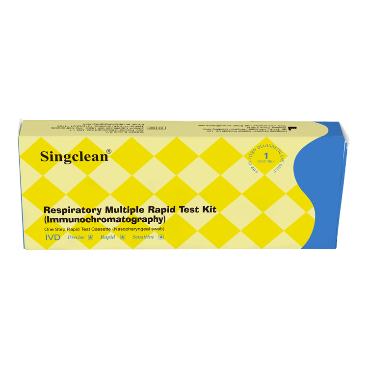
Respiratory Multiple Rapid Test Kit (Immunochromatography)
Instructions for use
For in vitro diagnostic use only
CE
INTENDED USE
Respiratory Multiple Rapid Test Kit (Immunochromatography) is used for in vitro qualitative detection of COVID-19 antigen, Respiratory Syncytial Virus antigen, Adenovirus antigen, MP antigen and influenza A/B antigen in human nasopharyngeal swab samples. This test provides only a preliminary test result. The test is suitable for all people with common cold symptoms or for common cold screening. This test is suitable for Professional Use only.
PACK FORMATS
1 Test/Box,5 Tests/Box ,10 Tests/Box,20 Tests/Box
INTRODUCTION
The novel coronaviruses belong to the β genus. COVID-19 is an acute respiratory infectious disease. People are generally susceptible. Currently, the patients infected by the novel coronavirus are the main source of infection; asymptomatically infected people can also be an infectious source. Based on the current epidemiological investigation, the incubation period
is 1 to 14 days, mostly 3 to 7 days.
Respiratory syncytial virus (RSV) belongs to the genus Pneumovirus of the family Paramyxoviridae. It can be infected by coughing and air droplets, mainly causing lower respiratory tract infections such as bronchiolitis and pneumonia in infants under 6 months, and upper respiratory tract infections such as rhinitis and cold in older children and adults, and bronchitis or pneumonia in the elderly.
Adenovirus infection can lead to respiratory tract, gastrointestinal tract, urinary tract and other related diseases, such as pharyngitis, pneumonia, conjunctivitis, cystitis and so on. Adenovirus infection has been a large-scale epidemic, can have adverse effects on the health of the population, causing fever, cough, sputum, throat congestion, eyes red, dry itching, vision loss, diarrhea, vomiting and other discomfort, respiratory symptoms are severe, can appear breathing difficulties, and even asphyxia and other serious consequences.
Mycoplasma pneumoniae (MP)can cause acute respiratory infection, clinical manifestations of headache, fever, dry cough, sore throat, muscle pain and other symptoms. All age groups are infected, but young and middle-aged people and children under 4 years of age have higher rates of infection. 30% of infected people will have a whole-lung infection. Mycoplasma pneumoniae can also cause non-respiratory diseases, such as meningitis, encephalitis, pancreatitis and so on.
Influenza viruses (Flu) are the pathogens that cause influenza. Influenza is an acute respiratory infection caused by influenza A, B, and C viruses. Influenza A virus often appears in epidemic form and can cause a worldwide influenza pandemic. Influenza B virus often causes local outbreaks and does not cause a worldwide influenza pandemic. Influenza A/B viruses (Flu A/B) can cause acute respiratory infections, and people are generally susceptible.
These viruses are highly contagious, spread quickly, have a short incubation period, and have a high incidence. The main symptoms are fever, dry cough, fatigue, etc. Therefore, the joint detection of COVID-19, RSV, ADV , MP and FLU A/B has relatively great clinical findings.
PRINCIPLE
These tests use colloidal gold immunochromatography to detect COVID-19, RSV, ADV , MP and FLU A/B antigens. The test of COVID-19 use COVID-19 monoclonal antibody (test line T) and goat anti-chicken polyclonal antibody (control line C) immobilised on a nitrocellulose strip. The burgundy colored conjugate pad contains colloidal gold conjugated to another COVID-19 monoclonal antibody. When the processed buffer containing the sample is added to the sample well, COVID-19 will combine with the COVID-19 monoclonal antibody conjugate to form an antigen-antibody complex. This complex migrates through nitrocellulose membrane by capillary action. When the complex meets the line of the COVID-19 monoclonal antibody of test line T, the complex is trapped forming a burgundy colored band which confirms a reactive test result. Absence of a colored band in the test means negative test result.
The test of RSV use RSV monoclonal antibody (test line T) and goat anti-chicken polyclonal antibody (control line C) immobilised on a nitrocellulose strip. The burgundy colored conjugate pad contains colloidal gold conjugated to another RSV monoclonal antibody. When the processed buffer containing the sample is added to the sample well, RSV will combine with the RSV monoclonal antibody conjugate to form an antigen-antibody complex. This complex migrates through nitrocellulose membrane by capillary action. When the complex meets the line of the RSV monoclonal antibody of test line T, the complex is trapped forming a burgundy colored band which confirms a reactive test result. Absence of a colored band in the test means negative test result
The test of ADV antigen use ADV monoclonal antibody (test line T) and goat anti-chicken polyclonal antibody (control line C) immobilised on a nitrocellulose strip. The burgundy colored conjugate pad contains colloidal gold conjugated to another ADV monoclonal antibody. When the processed buffer containing the sample is added to the sample well, ADV will combine with
the ADV monoclonal antibody conjugate to form an antigen-antibody complex. This complex migrates through nitrocellulose membrane by capillary action. When the complex meets the line of the ADV monoclonal antibody of test line T, the complex is trapped forming a burgundy colored band which confirms a reactive test result. Absence of a colored band in the test means
negative test result.
The test of Mycoplasma pneumoniae (MP) use MP monoclonal antibody (test line T) and goat anti-chicken polyclonal antibody (control line C) immobilised on a nitrocellulose strip. The burgundy colored conjugate pad contains colloidal gold conjugated to another MP monoclonal antibody. When the processed buffer containing the sample is added to the sample well, MP will combine with the MP monoclonal antibody conjugate to form an antigen-antibody complex. This complex migrates through nitrocellulose membrane by capillary action. When the complex meets the line of the MP monoclonal antibody of test line T, the complex is trapped forming a burgundy colored band which confirms a reactive test result. Absence of a colored band in the test means negative test result.
The test of Flu A/B is coated with influenza A virus monoclonal antibody (test line A), influenza B virus monoclonal antibody (test line B) and goat anti-chicken polyclonal antibody (control line C) on a nitrocellulose membrane. The gold label pad contains another influenza A virus monoclonal antibody and influenza B virus monoclonal antibody coupled with colloidal gold. When a sample containing influenza A virus antigen is added to the sample well, the influenza A virus antigen will combine with the influenza A virus monoclonal antibody conjugate to form an antigen-antibody complex. The complex moves through the nitrocellulose membrane. When the complex encounters the influenza A virus monoclonal antibody on the test line A, the complex is captured and forms a burgundy band, which confirms the positive test result.
When a sample containing influenza B virus antigen is added to the sample well, the influenza B virus antigen is the same as that of influenza A virus antigen. The influenza B virus antigen will color the test line B, forming a burgundy line, that is, confirm Influenza B virus positive test result. No color band on test line A/B means negative test result.
The test kit contains a quality control line (control line C). A purple-red line appears on the control line regardless of the
presence or absence of COVID-19, RSV, ADV , MP and FLU A/B Antigen in the sample. If the control line is not colored, it means that the test result is invalid regardless of whether the test line is colored. The control line is the criterion for judging whether the test strip is valid.
MATERIALS PROVIDED
1. Sealed pouches each containing a test cassette, a desiccant
2. Sampling cotton swab
3. Antigen Extraction Tube with Extraction Reagent
4. Instruction for use
MATERIALS REQUIRED BUT NOT PROVIDED
Timer
STORAGE AND STABILITY
The test kit can be stored at room temperature or refrigerated (4-30°C). The test device is stable through the expiration
date printed on the sealed pouch. The test device must remain in the sealed pouch until use.
Do not freeze.
Do not use beyond the expiration date.
After opening the sealed pouch, use the test as soon as possible within 60 minutes.
WARNINGS AND PRECAUTIONS
This test provides only a preliminary screening COVID-19, RSV, ADV , MP and FLU A/B Antigen test result. Therefore, any positive specimen with the Respiratory Multiple Rapid Test Kit (Immunochromatography) must be confirmed with more accurate testing methods and clinical results.
1.Do not use after expiration date.
2.All users have to read the instruction prior to performing a test. These instructions must be strictly followed.
3.Do not use it if the tube/pouch is damaged or broken.
4.Do not use if package is open or damaged.
5.Wear protective while handling specimens and wash hands thoroughly after wards.
6.Avoid splashing or aerosol formation of specimen and buffer.
7.Clean up spills thoroughly using an appropriate disinfectant.
8.Decontaminate and dispose of all specimens, reaction kits and potentially contaminated materials (i.e. swab, Antigen Extraction Tube with Extraction Reagent, test device) in a biohazard container as if they were infectious waste and dispose according to applicable local regulations.
9.Do not mix or interchange different specimens.
10.Do not mix reagent of different lots or those for other products.
11.Do not store the test kit in direct sunlight.
12.To avoid contamination, do not touch the head of provided swab when opening the swab pouch.
13.The provided swab in the package should be used only for nasopharyngeal specimen collection.
14.To avoid cross-contamination, do not reuse the swab for specimen collection.
15.Do not dilute the collected swab with any solution except for the provided extraction buffer.
16.Test is for single use only. Do not re-use under any circumstances.
17.Do not perform the test in a room with strong air flow, ie. electric fan or strong air-condition.
18.If the extracting solution touches eyes and mouth, a large amount of clean water should be rinsed in time and medical treatment should be sought if needed.
19.In case of any serious incident related to the device, it shall be reported to the manufacturer, distributor, and the competent authority.
SPECIMEN COLLECTION
1. Respiratory Multiple Rapid Test Kit (Immunochromatography)can be performed using nasopharyngeal swab.
2. The sample should be used as soon as possible after collection, otherwise it should be stored in a closed container at room temperature, but not more than 24 hours. Store at 2-8°C for one week. Long-term storage please refrigerate. Improper handling and storage of samples may result in false negative results.
3. If specimens are to be shipped, they should be packed in compliance with local regulations covering the transportation of etiologic agents.
TEST PROCEDURE
Allow test cassette, Antigen Extraction Tube with Extraction Reagent to room temperature (15-30°C) prior to testing.
1.Ask the patient to remove the secretions on the surface of the anterior nasopharynx, keep the head slightly tilted, and gently and slowly insert the swab through the nasopharynx to the nasopharynx, When resistance is encountered, it will reach the posterior nasopharynx, stay for a few seconds to absorb secretions, and gently rotate to remove the swab.

2. Put the swab specimen into the Antigen Extraction Tube with Extraction Reagent, and rotate the swab about 10 times while pressing the swab head against the tube wall to release the antigen in the swab, then let it stand for about 1 minute.
3. Remove the swab while squeezing the tip of the swab so that as much liquid in the swab can be discharged as possible. Dispose of used swabs in accordance with biohazard waste disposal methods.
4. Install the dripper on the Antigen Extraction Tube with Extraction Reagent and cap it tightly, and let it stand for about 1 minute.
5.Open the aluminum foil bag and take out the test card, add 3 drops (about 100μL) into the sample hole of the test card (or use a pipette to add 100μL), and start the timer.
6. Wait for the colored line to appear. The result should be read after 15 minutes. Do not interpret the result after 20 minutes.

INTERPRETATION OF RESULTS
For COVID-19 Antigen test kit:
NEGATIVE:
If only the C band is present, the absence of any burgundy color in the T band indicates that no COVID-19 antigen is detected in the specimen. The result is negative.
POSITIVE:
In addition to the presence of C band, if T band is developed, the test indicates for the presence of COVID-19 antigen in the specimen. The result is COVID-19 positive.
INVALID:
Control line fails to appear. Insufficient specimen volume or incorrect procedural techniques are the most likely reasons for control line failure. Review the procedure and repeat the test with a new test cassette. If the problem persists, discontinue using the test kit immediately and contact your local distributor.
For RSV Antigen test kit:
NEGATIVE:
If only the C band is present, the absence of any burgundy color in the T band indicates that no RSV antigen is detected in the specimen. The result is negative.
POSITIVE:
In addition to the presence of C band, if T band is developed, the test indicates for the presence of RSV antigen in the specimen. The result is RSV positive.
INVALID:
Control line fails to appear. Insufficient specimen volume or incorrect procedural techniques are the most likely reasons for control line failure. Review the procedure and repeat the test with a new test cassette. If the problem persists, discontinue using the test kit immediately and contact your local distributor.
For ADV Antigen test kit:
NEGATIVE:
If only the C band is present, the absence of any burgundy color in the T band indicates that no ADV antigen is detected in the specimen. The result is negative.
POSITIVE:
In addition to the presence of C band, if T band is developed, the test indicates for the presence of ADV antigen in the specimen. The result is ADV positive.
INVALID:
Control line fails to appear. Insufficient specimen volume or incorrect procedural techniques are the most likely reasons for control line failure. Review the procedure and repeat the test with a new test cassette. If the problem persists, discontinue using the test kit immediately and contact your local distributor.
For MP Antigen test kit:
NEGATIVE:
If only the C band is present, the absence of any burgundy color in the T band indicates that no MP antigen is detected in the specimen. The result is negative.
POSITIVE:
In addition to the presence of C band, if T band is developed, the test indicates for the presence of MP antigen in the specimen. The result is MP positive.
INVALID:
Control line fails to appear. Insufficient specimen volume or incorrect procedural techniques are the most likely reasons for control line failure. Review the procedure and repeat the test with a new test cassette. If the problem persists, discontinue using the test kit immediately and contact your local distributor.
For Flu A/B Antigen test kit:
NEGATIVE:
If only the C band is present, the absence of any burgundy color in the T band indicates that no Flu A/B antigen is detected in the specimen. The result is negative.
POSITIVE
FLU A and FLU B positive:
In addition to the presence of the C-line, If the test lines A and B appears at the same time, it means that there are both influenza A virus antigen and influenza B virus antigen in the sample, that is, the result is positive for FLU A and FLU B.
FLU A positive:
In addition to the presence of the C-line, if the test line A appears, the test indicates the presence of FLU A antigen in the sample, that is, the result is positive for FLU A.
FLU B positive:
In addition to the presence of the C-line, if the test line B appears, the test indicates the presence of FLU B antigen in the sample, that is, the result is positive for FLU B.
INVALID:
Control line fails to appear. Insufficient specimen volume or incorrect procedural techniques are the most likely reasons for control line failure. Review the procedure and repeat the test with a new test cassette. If the problem persists, discontinue using the test kit immediately and contact your local distributor.